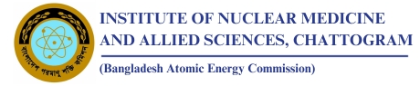
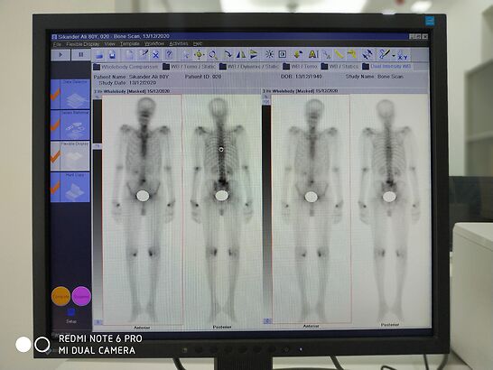
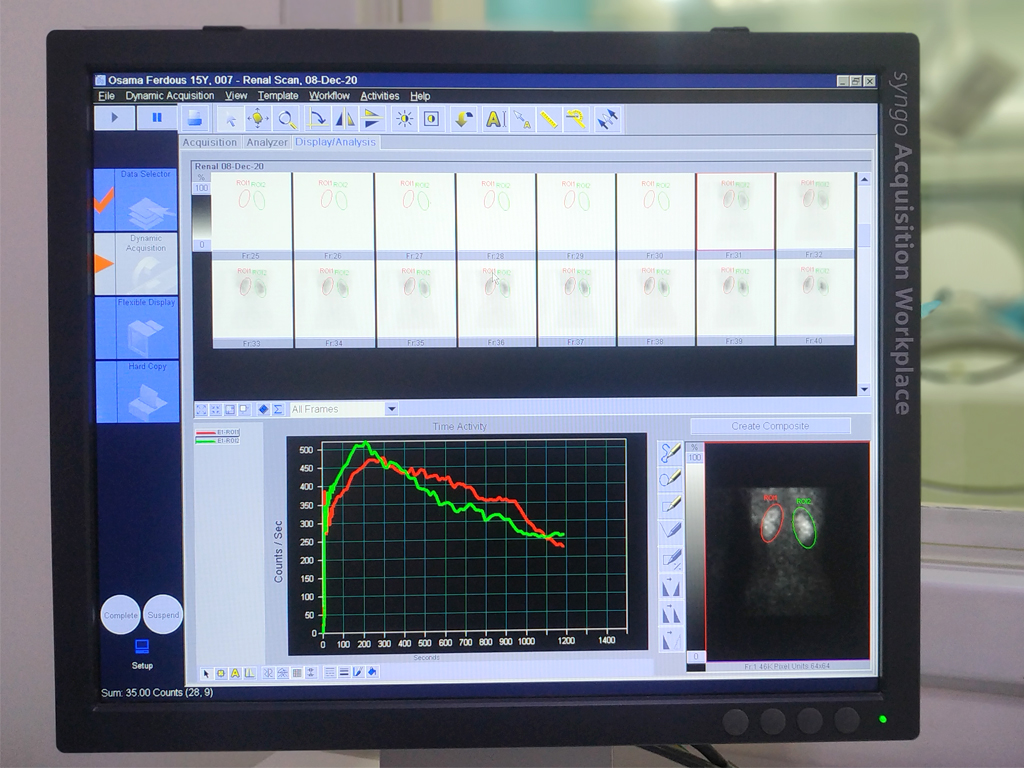
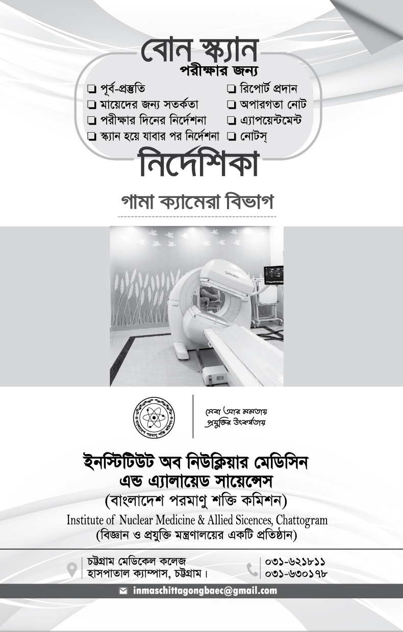
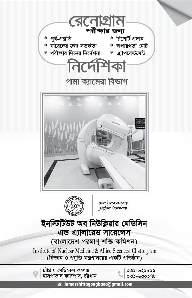
Gamma Camera: The special camera and imaging techniques used in nuclear medicine include the Gamma camera and single-photon emission computed tomography (SPECT). The Gamma camera, also called a scintillation camera, detects radioactive energy that is emitted from the patient's body and converts it into an image. It simultaneously detects radiation from the entire field of view and enables the acquisition of dynamic as well as static images of the area of interest in the human body.
Nuclear medicine imaging uses small amounts of radioactive material to diagnose, evaluate or treat a variety of diseases. These include many types of cancers, heart disease, gastrointestinal, endocrine or neurological disorders and other abnormalities. Because nuclear medicine exams can pinpoint molecular activity, they have the potential to identify disease in its earliest stages. They can also show whether a patient is responding to treatment. Nuclear medicine imaging procedures are non-invasive. With the exception of intravenous injections, they are usually painless.
Depending on the type of exam, the radiotracer is injected, swallowed or inhaled as a gas. It eventually accumulates in the area of the body under examination. A special camera or imaging device detects radioactive emissions from the radiotracer. The camera or device produces pictures and provides molecular information.
Many centers superimpose nuclear medicine images with computed tomography (CT) or magnetic resonance imaging (MRI) to produce special views. This is known as image fusion or co-registration. These views allow the doctor to correlate and interpret information from two different exams on one image this leads to more precise information and accurate diagnoses. Single photon emission computed tomography/computed tomography (SPECT/CT) and positron emission tomography/computed tomography (PET/CT) units can perform both exams at the same time.
Every Saturday to Thusday at 8.00am -2.30pm
Except Friday and all government holiday
Every Saturday to Thusday at 7.30 AM
Except Friday and all government holiday
| Invastigations | Rate | Preparation |
|---|---|---|
| DTPA-Renogram and Split Renal Function (Tc-99m) | 3000 | Get Appointment |
| Thyroid Scan (Tc-99m) | 500 | Get Appointment |
| Brain Scan (Tc-99m) | 600 | Get Appointment |
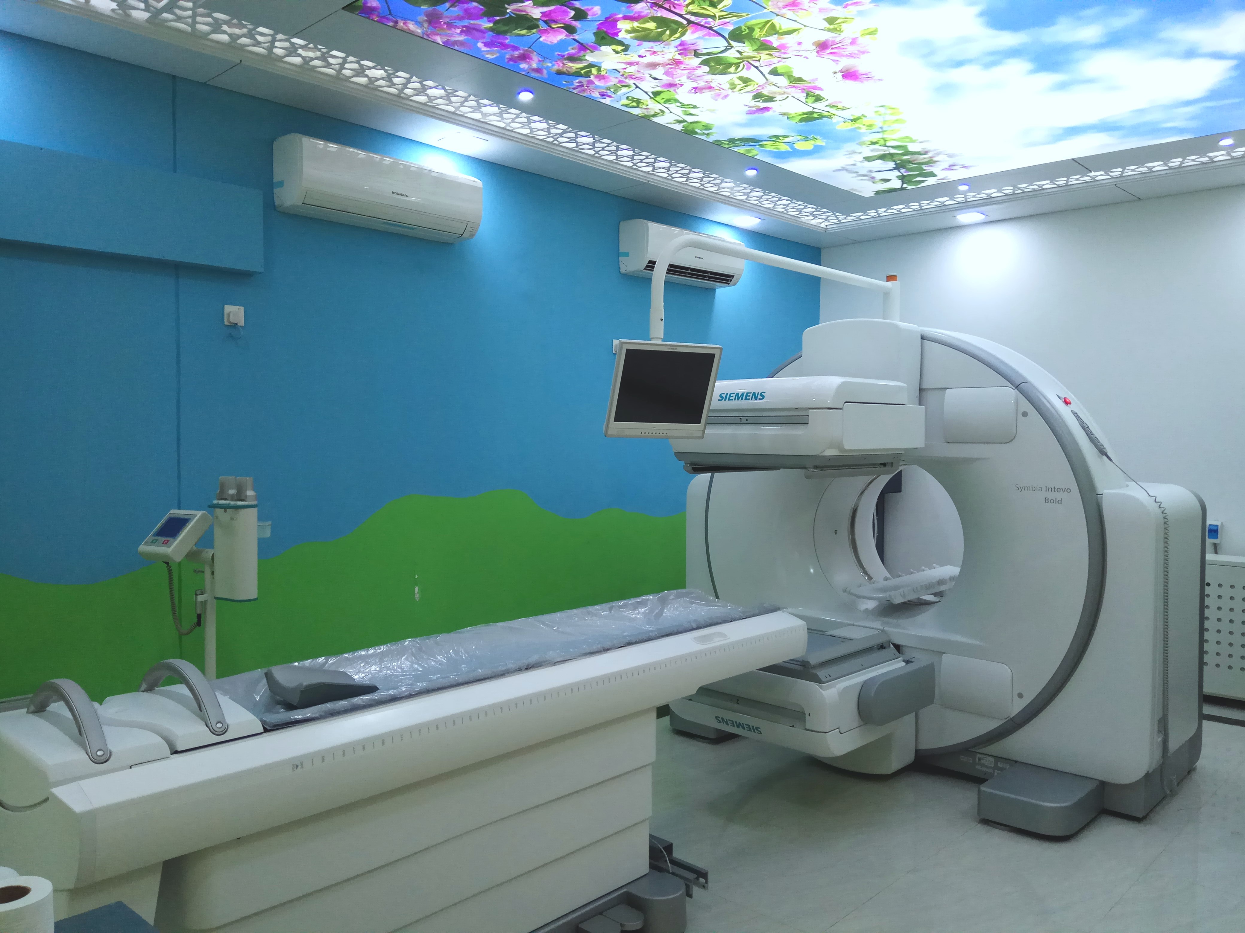
| 3 Phase Bone Scan | 3000 | Get Appointment |
| SPECT: Bone Scan | 4000 | Get Appointment |
| Kidney Scan (DTPA) | 1000 | Get Appointment |
| DMSA - Renal Scan (Tc 99m) | 3000 | Get Appointment |
| DTPA - Captopril Gamma Camera Renogram (Tc 99m) | 3000 | Get Appointment |
| Myocardial Perfusion Scan (MPI) | 7000 | Get Appointment |
| Cardiac MUGA | 4500 | Get Appointment |
| Carcinoid Tumor Evaluation With Octreotide | 20000 | Get Appointment |
| CSF Rhinorrhea Study | 3000 | Get Appointment |
| DTPA-Renogram and Serum Sample GFR (Tc 99m) | 3000 | Get Appointment |
| DTPA-Renogram With Camera GFR (Tc 99m) | 3000 | Get Appointment |
| GIT Studies (Gastric Emptying, Oesophageal Reflux) | 4000 | Get Appointment |
| Hepatobilliary Scan (Tc 99m) | 3000 | Get Appointment |
| Liver Perfusion/Flow | 3000 | Get Appointment |
| Hysterosalphingo Scintigraphy | 3000 | Get Appointment |
| Liver Scan (Tc 99m) | 3000 | Get Appointment |
| Liver Spleen Scan (Tc 99m) | 3000 | Get Appointment |
| Lymphoscintigraphy For Lymphatic Drainage Evaluation | 4000 | Get Appointment |
| Lymphoscintigraphy For Sentinel LM (Tc 99m) | 4000 | Get Appointment |
| Meckels Diverticulum Scan | 3000 | Get Appointment |
| MIBG Scan | 20000 | Get Appointment |
| Onchological Study MIBI, Thallium or Gallium | 20000 | Get Appointment |
| RBC - Scan for Hemangioma | 3000 | Get Appointment |
| Salivary Gland Scan | 3000 | Get Appointment |
| Scan for Gastro - Intestinal Bleeding (RBC) | 3000 | Get Appointment |
| Single Spot Bone Scan | 3000 | Get Appointment |
| Tc 99m PSMA | 8000 | Get Appointment |
| Testicular Scan | 3000 | Get Appointment |
| V - P Shaunt Patency Study | 3000 | Get Appointment |
| Vesicoureteric Reflux Study | 3000 | Get Appointment |
| SPECT: Kidney Scan | 4000 | Get Appointment |
| SPECT: Liver Scan | 4000 | Get Appointment |
| SPECT: Lung Perfusion | 4000 | Get Appointment |
| SPECT: Lung VQ Scan | 4000 | Get Appointment |
| SPECT MIBI parathyroid imaging | 4000 | Get Appointment |
| SPECT Myocardial perfusion (rest) | 4000 | Get Appointment |
| SPECT Myocardial perfusion (stress+ rest) | 10000 | Get Appointment |
| SPECT pyp Cardiac Scan | 5000 | Get Appointment |
| SPECT-CT Brain Scan | 5000 | Get Appointment |
| SPECT-CT Brain Tumor Recurrence | 5000 | Get Appointment |
| SPECT-CT Liver Spleen Scan | 5000 | Get Appointment |
| SPECT-CT Lung VQ Scan | 5000 | Get Appointment |
| SPECT-CT Other | 5000 | Get Appointment |
| SPECT-CT Parathyroid | 5000 | Get Appointment |
| SPECT-CT Salivary Scan | 5000 | Get Appointment |
| SPECT-CT Whole body bone scan | 5000 | Get Appointment |
| SPECT HMPAO Cerebral Perfusion Imaging (Tc99m) | 4000 | Get Appointment |
Common Clinical Applications
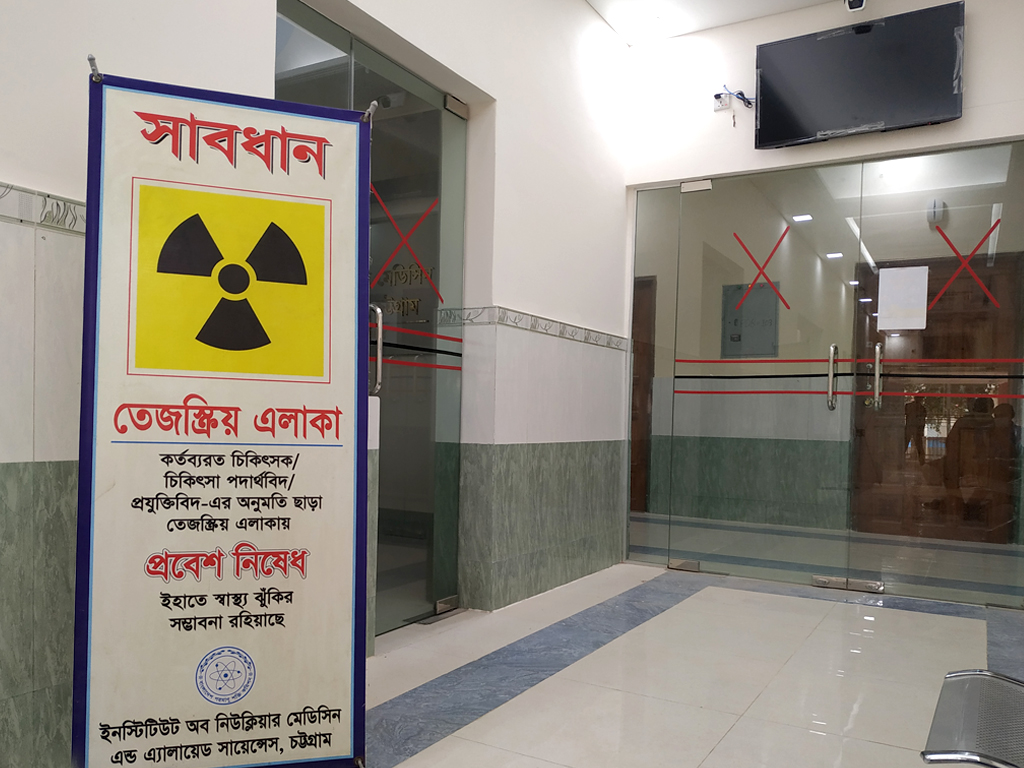
Safety
Bone Scan PDF/Print
Renogram PDF/Print
Regrets
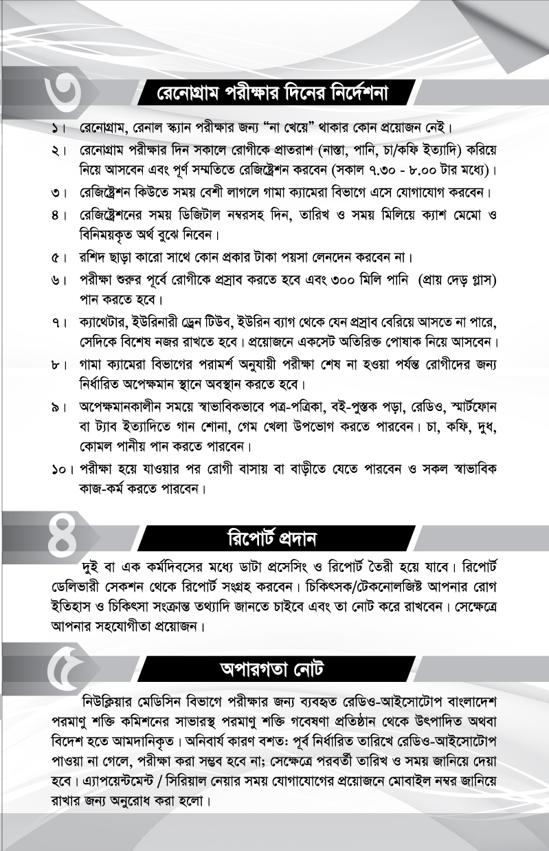
The ‘radio-isotope’ used for testing in the Nuclear Medicine is produced from the Atomic Energy Research Establishment at Savar, Dhaka of Bangladesh Atomic Energy Commission or imported. The uncertain reason is that, if the radio-isotope is not available at a pre-determined date, it will not be possible to do radio-isotope scan test for the patient; the date and time will be reported later. When making an appointment/serial, we need your mobile number to contact you later for any requirements.
Contact us now to schedule an appointment.